Welcome to Xi’an Good Doctor Medical Science and Technology Co., Ltd.website !
Internet Drug Information Service Qualification Certificate: (Shaanxi)-Non-Operating -2023-0143


How to distinguish between cancerous ascites and non-cancerous ascites
The abdominal cavity of a normal person contains only a small amount of fluid, which serves to lubricate the internal organs. When the volume of fluid in the abdominal cavity exceeds 200 ml, it is referred to as ascites. This article elaborates on the diagnostic characteristics and treatment methods for different types of ascites, aiming to help more clinicians better identify and address this clinical issue.
2024-09-12
The normal abdominal cavity contains only a small amount of fluid, which lubricates the internal organs. When the amount of fluid in the abdominal cavity exceeds 200 ml, it is called ascites. This article elaborates on the diagnostic characteristics and treatment methods for different types of ascites, aiming to help more clinicians better identify and address this clinical issue.
Characteristics of ascites caused by different etiologies
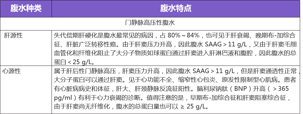
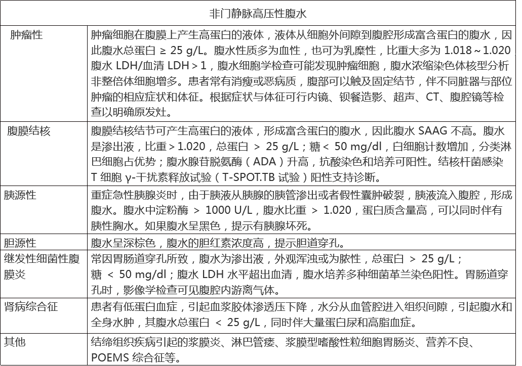
The causes of ascites can be divided into hepatic, cardiac, cancerous, vascular, renal, malnutrition-related, and tuberculous, making the diagnosis and differentiation of ascites causes very important. Diagnostic puncture and analysis of ascites is one of the most important steps.Laparoscopic examination with biopsy is the "gold standard" for diagnosing the cause of ascites., for unexplained ascites, this examination should be considered when the patient's condition allows.
Table 1. Characteristics of ascites caused by different etiologies
Biochemical testing related to the diagnosis of benign and malignant ascites[1]
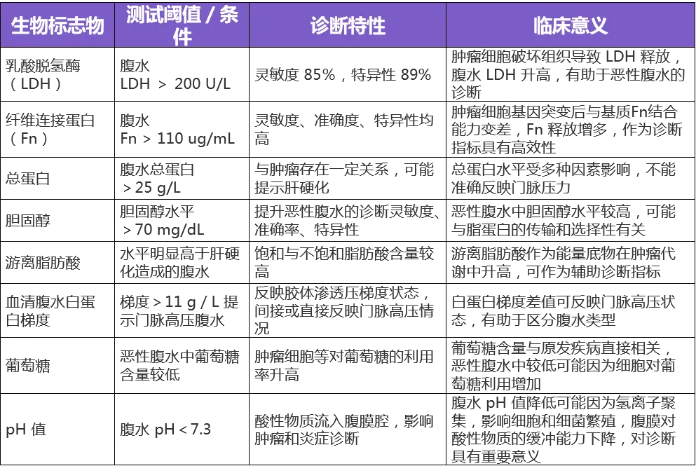
There are many biochemical items for clinical detection of ascites, among whichthe common biochemical indicators with significant clinical application arebriefly described as follows.
Table 2. Biochemical testing related to the diagnosis of benign and malignant ascites
Strategies for managing cancerous ascites
Clinically, ascites can be classified into grade 1 (small), grade 2 (moderate), and grade 3 (large) based on the volume of ascites.
▌Grade 1 or small ascites:Ascites that can only be detected by ultrasound, with patients generally showing no signs of abdominal distension, and negative shifting dullness; ultrasound ascites depth < 3 cm.
▌Grade 2 or moderate ascites:Patients often have moderate abdominal distension and symmetrical abdominal bulging, with shifting dullness negative/positive; ultrasound ascites depth 3-10 cm.
▌Grade 3 or large ascites:Patients have significant abdominal distension, positive shifting dullness, and may have abdominal bulging or even umbilical hernia formation; ultrasound ascites depth > 10 cm.[2]
Treatment targeting the cancer itself is the fundamental method to reduce ascites. Controlling tumor growth and spread through chemotherapy, radiotherapy, targeted therapy, etc., helps reduce the production of ascites. At the same time, for specific types of cancer, such as liver cancer and ovarian cancer, using targeted treatment plans can more effectively control ascites. Based on systemic treatment, local treatment can be used to alleviate symptoms.
Table 3. Summary of methods for malignant ascites

References
[1] Chen Yang. Progress in biochemical testing related to the diagnosis of benign and malignant ascites [J]. China Urban and Rural Enterprise Health, 2024, 39(05): 43-45.
[2] ChineseMedical Association Hepatology Branch. Guidelines for the diagnosis and treatment of cirrhotic ascites [J]. Chinese Journal of Hepatology, 2023, 31(8): 813-826.
Source: Dingxiangyuan Oncology Time, content is for sharing purposes only, please delete if infringing.
Key words:
Related News

· China Good Doctor ·
· Good friends of human beings ·
Address: Floor 1-2, Left Building No. 1, Huoju Road, East zone of Xi’an High- tech Development District, Xi’an, China
Copyright Xi'an Good Doctor Medical Technology Co., Ltd.
Internet Drug Information Service Qualification Certificate (Shaanxi)-Non-Operating -2023-0143